Masked AAV Vectors Make Gene Delivery Respond to Tissue, Light, or Injury
A Nature Materials study in mice adds an activation step to AAV gene delivery: the vector stays masked until a liver enzyme, near-infrared light, or injury-related chemistry removes the block. The result is promising targeting research, not a ready human treatment.

Adeno-associated virus, or AAV, has become one of the main delivery vehicles for gene therapy because it can carry genetic instructions into cells without behaving like a disease-causing virus. Its usefulness has always come with an awkward trade-off: once injected, the vector can reach more cells and tissues than clinicians would ideally want. A dose that travels broadly may also expose the liver, muscles, brain, heart, or immune system to material intended for only one location.
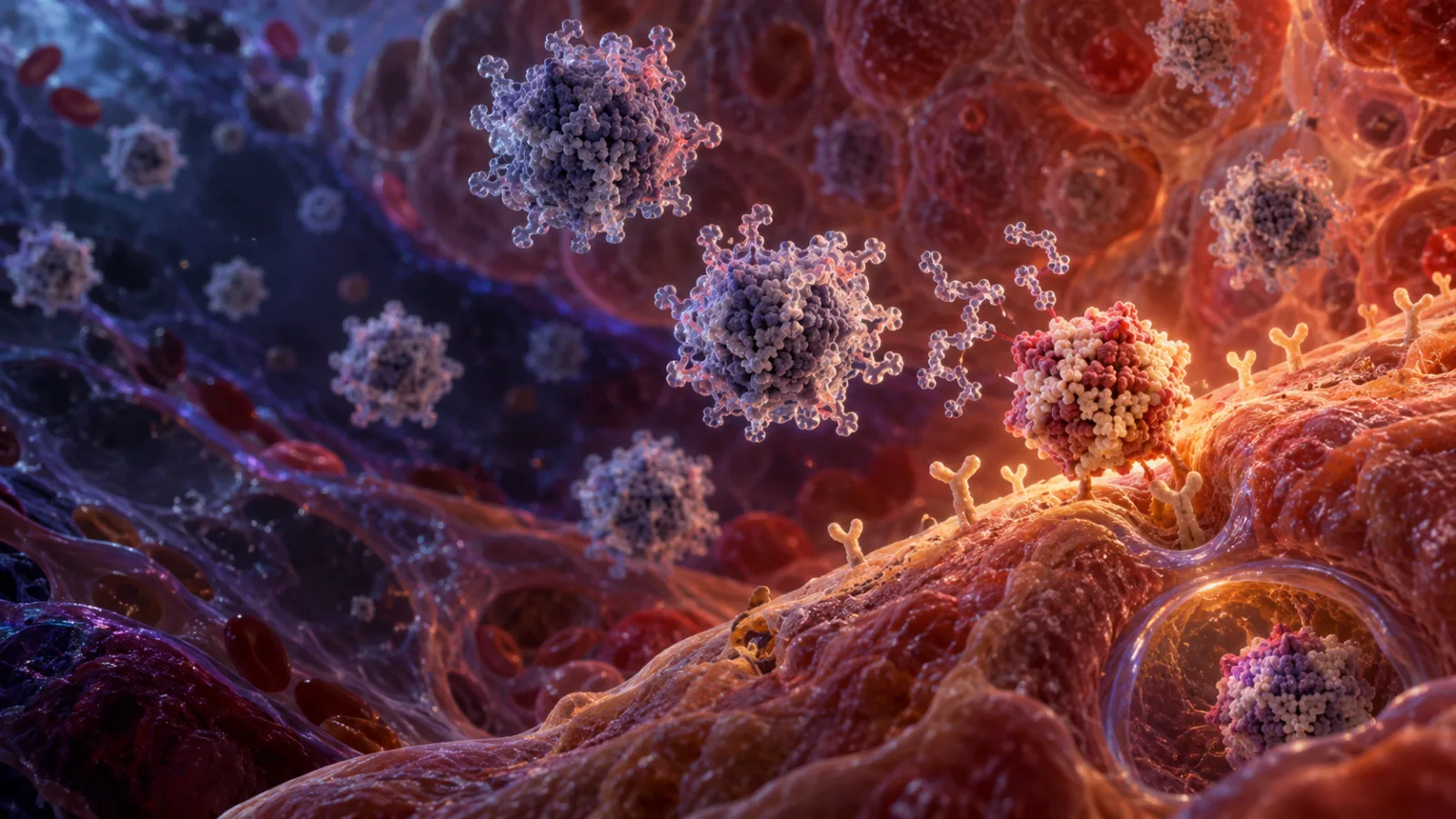
A study published in Nature Materials on 29 September 2026 reports a way to add a second layer of control. The researchers built AAV particles whose ability to enter cells is chemically blocked at first. The masking group can then be removed by a local biological signal or by an external near-infrared-light trigger. In mice, the approach directed gene delivery toward the liver, illuminated muscle and brain regions, and injured heart tissue.
The important advance is not that AAV can carry a gene. That has been demonstrated repeatedly, including in approved therapies. The advance is the attempt to make the vector wait for permission from the tissue before it becomes effective. It addresses one of the hardest practical questions in in-vivo gene therapy: how to deliver enough cargo to the intended cells while reducing exposure elsewhere.
Why AAV targeting remains difficult
AAV is a small viral vector assembled from a protein shell, or capsid, surrounding genetic material. Different AAV serotypes and engineered capsids show different preferences for tissues, but “preference” is not the same as exclusivity. A vector selected for liver delivery can still circulate through other organs. A vector designed to reach the nervous system may encounter barriers, immune defenses, and unintended cell types along the way.
The problem becomes more serious when the vector is administered systemically. Intravenous delivery is attractive because it can reach tissues that are difficult to access directly, but the whole body sees the particle before it reaches its destination. The amount of vector required to obtain useful expression in a hard-to-reach organ can increase exposure in organs that act as sinks, especially the liver.
There is also an immune constraint. The US Food and Drug Administration notes that AAV capsids can trigger antibody and cellular immune responses, and that more than 90% of people have binding antibodies to some AAV serotypes. Some of those antibodies neutralize the vector before it can enter cells. Immune responses can reduce efficacy, eliminate transduced cells, or make repeat dosing difficult. These are not minor engineering details; they affect whether a therapy can be given, how much vector is needed, and whether it can be used again later.
The new study does not remove those challenges. It changes the first encounter between the vector and the body. Instead of relying only on capsid tropism, the researchers temporarily cover the capsid’s ability to interact with cellular receptors. The vector is designed to circulate in a less active state, then regain infectivity where a chosen trigger removes the cover.
The masking system in plain language
The team used genetic-code expansion to place a non-standard amino acid containing a tetrazine group into selected AAV capsid proteins. That chemical handle allowed the researchers to attach a blocking structure to the particle. The block interfered with the interaction needed for AAV to infect a cell.
The researchers tested two broad ways to create the blocked state. In one, the modified capsid was connected to a truncated form of the AAV receptor. In another, polyethylene glycol, commonly called PEG, was attached to the capsid. Both strategies were intended to reduce or stop transduction until a cleavable linker was removed.
The design then paired the masked particle with a trigger. The study describes three types of activation:
- A liver-specific protease called matriptase-2, also known as TMPRSS6, could remove a masking group in the liver.
- Near-infrared light could activate particles in illuminated regions, including selected muscle and brain areas.
- Reactive oxygen species associated with tissue damage could activate particles in an injured heart model.
This is more precise than simply choosing a different AAV serotype, but it is not a universal address system. Each trigger has its own biological range, depth, timing, and risk of unintended activation. The useful idea is the separation of delivery from activation: a particle can be present in the body without being fully competent to enter cells until a second condition is met.
What the mouse experiments showed
In the liver experiment, the researchers administered a protease-activated masked AAV systemically. Because matriptase-2 is associated with liver biology, the masking group could be removed preferentially there. The paper reports tissue-selective gene delivery in mice rather than a general claim that all liver cells were reached and all other organs were untouched. That distinction matters. Biological targeting is usually a matter of distribution and relative enrichment, not a perfect on-off switch.
The light-activated version used near-infrared illumination as the external control. Near-infrared light is more useful than visible light for tissue experiments because it can penetrate farther into biological material, although penetration remains limited and depends on wavelength, tissue composition, power, and geometry. In the study, illumination restored AAV activity in selected regions such as muscle and brain.
The brain result is best understood as a proof of localized activation in an animal model, not as evidence that an intravenous treatment can now be aimed at any human brain structure from outside the skull. Mouse anatomy is smaller, experimental lighting can be carefully arranged, and the delivery conditions do not reproduce the full complexity of a clinical procedure.
The third design used inflammation-associated reactive oxygen species as a disease-linked signal. In a mouse model of cardiac ischaemia-reperfusion injury, the activated vector delivered a gene encoding VEGF-A165 to the myocardium. The authors report localized expression and improved cardiac function in that model. VEGF-A is involved in blood-vessel growth, so the experiment tested whether injury-responsive delivery could support repair in damaged heart tissue.
That result is encouraging for a specific reason: the trigger was tied to the condition being treated rather than to an arbitrary anatomical landmark. A vector that becomes active in an injured environment could, in principle, reduce expression in healthy tissue. But the present evidence remains preclinical. A mouse model of ischaemia-reperfusion injury is not the same as chronic human heart disease, and a change in cardiac measurements is not proof of durable clinical benefit.
Why this is different from ordinary tissue tropism
Traditional AAV engineering often asks which capsid naturally enters a target tissue most efficiently. Researchers can also alter the capsid surface, attach targeting ligands, change the promoter that controls gene expression, or administer the vector directly to an organ. Each method acts at a different stage.
Capsid tropism influences where the particle goes and which cells it can enter. A promoter influences which cells produce the encoded protein after the vector has delivered its genetic payload. Direct administration changes the route and concentration. The masked-vector approach adds a gate before cell entry: the particle is designed to remain less infectious until a trigger removes the barrier.
That extra gate could be useful when a therapeutic gene is powerful enough that off-target expression matters. It could also reduce the amount of active vector encountered by non-target tissues, although the study does not establish that this will reduce clinical toxicity in humans. The practical question is not simply whether masking works in a controlled experiment. It is whether enough particles can be produced consistently, remain stable in storage and blood, activate at the desired location, and avoid becoming active at the wrong time.
The strategy also fits a broader direction in gene-therapy research: control is being distributed across several layers instead of being assigned to one component. Scientists are working on capsids that prefer particular tissues, transient editors that do not remain active indefinitely, particles that release cargo under specific conditions, and gene circuits that respond to cellular state. A masked AAV is one contribution to that larger engineering problem.
The technical limitations are substantial
The first limitation is the model. The reported results are in mice. Mouse experiments are valuable for testing biodistribution, activation chemistry, and initial safety signals, but they do not predict human performance reliably enough on their own. Human tissues are larger, human immune histories are different, and the enzymes and disease environments used as triggers may vary from patient to patient.
The second limitation is trigger control. A liver enzyme may be relatively enriched in the intended organ without being exclusive to it. Reactive oxygen species are part of normal signaling as well as injury biology. If the activation threshold is too low, a particle could become active in places experiencing ordinary oxidative stress. If it is too high, the vector may fail to activate in diseased tissue. The therapeutic window must be measured rather than assumed.
Light creates a different set of constraints. Near-infrared illumination can provide spatial control at accessible depths, but light intensity falls as it travels through tissue and is affected by scattering and absorption. Reaching the deep human brain or a large internal organ would require a clinically acceptable method for delivering enough light to the right volume. The study demonstrates a controllable laboratory trigger; it does not solve the clinical hardware and dosimetry problem.
The chemical masking itself must also be scrutinized. Adding non-standard amino acids, linkers, receptor fragments, or PEG changes the surface of the vector. Those modifications could alter manufacturing yield, aggregation, circulation time, biodistribution, or recognition by the immune system. A particle that is less infectious before activation may still be visible to antibodies and immune cells. Masking receptor interaction is not the same as making the vector immunologically invisible.
AAV’s packaging capacity remains relevant as well. The vector can carry only a limited amount of genetic material, and adding control components or more elaborate expression systems competes with the therapeutic payload. The masking chemistry is applied to the capsid rather than inserted as a large genetic cassette, which helps, but the full therapeutic product would still have to fit within AAV’s manufacturing and packaging constraints.
Repeat dosing is another unresolved issue. After exposure, the body can develop antibodies against the capsid. A masked particle may change when and where it becomes infectious, but it is still built from AAV proteins. If the immune system recognizes the capsid before activation, later doses could remain difficult. The new system should therefore be viewed as a targeting intervention, not as a complete answer to AAV immunogenicity.
Finally, the study reports delivery and repair-related outcomes in animals, not long-term human safety. Gene delivery can persist, and the biological effect of the encoded protein may outlast the original trigger. A vector activated in a brief injury window could potentially produce a lasting change in the treated cells. That may be desirable, but it means that activation timing, expression duration, dose, and reversibility all need separate evaluation.
What would have to happen before clinical use
A credible path toward human testing would require more than reproducing a bright fluorescence signal in mouse tissue. Researchers would need to establish the amount of masked vector that reaches each organ, the fraction that remains inactive, the fraction that activates, and the duration of the active state. Those measurements would need to be repeated across sexes, ages, disease states, and relevant animal species.
The safety package would also have to examine immune responses to the capsid and the added chemical components, liver and kidney function, inflammatory markers, blood-clotting effects, tissue pathology, and the possibility of unwanted gene expression. For the heart application, it would be important to distinguish improved function caused by localized expression from effects caused by the delivery system itself or by the experimental injury model.
Manufacturing is a quiet but decisive test. The final product would need a defined ratio of masked and unmasked particles, a reliable level of capsid modification, low amounts of empty or damaged particles, and a shelf life suitable for clinical use. A trigger system that works only when every particle has the same chemical state will be difficult to translate if production creates a mixed population.
The field would also need a practical answer to patient selection. A protease-activated design depends on the target enzyme being present at the right level. An injury-activated design depends on the disease producing the required chemical environment. A light-activated design depends on access to the target tissue. These conditions may differ widely among patients, which could push such therapies toward highly specified indications rather than broad, one-size-fits-all treatment.
Regulators would likely ask a simple but demanding question: what happens when the trigger is present outside the intended tissue? The answer has to come from quantitative biodistribution and toxicology, not from the label “targeted.” The FDA’s discussion of AAV immunogenicity is a reminder that delivery vectors are themselves active biological products with their own safety profile.
The useful advance is control, not a finished therapy
The most defensible interpretation of this work is that it improves the control architecture of AAV gene delivery. The vector is no longer asked to solve every problem through capsid choice alone. It can be designed to remain masked, then activated by a tissue enzyme, an external light signal, or a disease-associated chemical environment. That gives researchers more knobs to tune.
The study does not show that gene therapy can now be directed perfectly to any organ. It does not demonstrate a human treatment, remove pre-existing immunity, guarantee repeat dosing, or prove that the heart-repair result will translate to patients. Those limits are not reasons to dismiss the result. They define what the next experiments need to measure.
Good technology news often arrives as a narrower claim than the headline version. Here, the claim is that AAV particles can be engineered to carry a reversible-looking activation barrier and that several triggers can restore delivery in selected mouse tissues. If later work confirms reliable activation, acceptable safety, scalable manufacturing, and useful performance in larger animals, masked vectors could become one part of a more precise gene-therapy toolkit. For now, the achievement is a meaningful step toward making delivery conditional on where and when the body gives the signal.
Related articles

AWS’s TOLAP release puts object-level access control inside AI-agent tools
AWS Labs’ TOLAP project treats the tool that fetches data—not just the agent identity or API gateway—as the enforcement point for rows, fields, actions, delegation and result limits.


Notification settings
Verify contacts, choose a device, event and available channel, understand immediate field saving and diagnose recognition separately from delivery.


My Eicher shows why connected fleet security starts with the API
A published fleet-platform case study points to account, GPS and document exposure risks without needing Hollywood claims about remote truck control.


Perplexity v. Amazon: the practical line between user-directed AI agents and bots
A U.S. appeals ruling narrows one anti-hacking argument against AI browser agents, but practical adoption still depends on consent, identity, logs, data minimization and platform-aware safety.

Comments
Sign in to comment.
No comments yet.